Advertisement

We need your help now

Support from readers like you keeps The Journal open.

You are visiting us because we have something you value. Independent, unbiased news that tells the truth. Advertising revenue goes some way to support our mission, but this year it has not been enough.

If you've seen value in our reporting, please contribute what you can, so we can continue to produce accurate and meaningful journalism. For everyone who needs it.
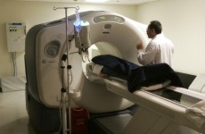
File photo of nuclear medicine technologist Michael Gomex speaking to a patient Mike Groll/AP/Press Association Images
Cancer

Childhood CT scans 'could triple leukaemia and brain cancer risk'

A study at Newcastle University showed that CT scans in children could triple their risk of contracting brain cancer or leukaemia when older. This is the first long-term study of its kind.

A LONG-TERM study has shown that radiation exposure from CT scans in childhood could triple a child’s risk of later developing brain cancer or leukaemia.

The study is discussed in an article in The Lancet, written by Dr Mark Pearce and Professor Sir Alan Craft, Newcastle University, UK; Professor Louise Parker, Dalhousie University, Halifax, NS, Canada;  Dr Amy Berrington de González, National Cancer Institute (NCI), National Institutes of Health (NIH), Bethesda, MD, USA, and colleagues.

The study represents the culmination of almost two decades of research in this area, and is jointly funded by the UK Department of Health and NCI/NIH.

CT scans

The authors say that CT – or computed tomography – scans are a vital diagnostic technique.

However their study shows that radiation exposure from two to three of them during childhood (under the age of 15) can triple the risk of later developing brain cancer.

Receiving five to 10 such scans could in turn triple the risk of developing leukaemia.

The scientists said that while the “absolute risk” of these cancers occurring after CT is “small”, radiation doses from CT scans should be kept as low as possible and alternative procedures that do not use ionising raidation “should be considered if appropriate”.

The potential cancer risks are linked to the ionising radiation used in CT scans.

Data

The study involved almost 180,000 patients who underwent a CT scan between 1985 and 2002 according to reports from the radiology departments of 70 per cent of the UK’s hospitals. The researchers took the number and types of CT scan and estimated the dose of radiation absorbed by the brain and bone marrow in patients for each scan.

They then linked the data to cancer incidence and mortality reports in the UK National Health Service Registry between 1985 – 2008, from which they calculated the excess incidence of leukaemia and brain tumours.

A total of 74 from 178, 604 patients were diagnosed with leukaemia and 135 of 176, 587 were diagnosed with brain cancer.

Risks

The authors calculated that the relative risk of leukaemia increased by 0.036 per extra mGy received, whilst for brain tumours this increased risk was 0.023.

Compared with patients who received a dose of radiation of less than 5 mGy, patients who received a cumulative dose of at least 30 mGy (mean 50 mGy) had around three times the risk of leukaemia.

Meanwhile, patients receiving a cumulative dose of 50-74 mGy (mean 60 mGy) had triple the risk of developing primary brain tumours.

The authors say that, of every 10, 000 people between the ages of 0-20 years receiving 10 mGy from a CT scan, there would be about one expected excess leukaemia case, whereas there were would be one excess case of brain cancer for every 30,000 people.

Applying the dose estimates for one head CT scan before the age of 10 years, this would translate into approximately one excess case of leukaemia and one excess brain tumour per 10, 000 patients in the decade after first exposure.

The authors note that increased follow-up and analysis of other cancer types is needed to identify the total excess risk for all cancers associated with CT scans.

Benefits and risks

Lead author Dr Pearce said that the immediate benefits of CT outweigh the potential long­term risks in many settings.

Further refinements to allow reduction in CT doses should be a priority, not only for the radiology community, but also for manufacturers. Alternative diagnostic procedures that do not involve ionising radiation exposure, such as ultrasound and MRI [magnetic resonance imaging] might be appropriate in some clinical settings. Of utmost importance is that where CT is used, it is only used where fully justified from a clinical perspective.

Dr Berrington de González added that it is well known that radiation can cause cancer, “but there is an ongoing scientific debate about whether relatively low doses of radiation like those received from CT scans do increase cancer risks, and if so the magnitude of those risks”.

Ours is the first study to provide direct evidence of a link between exposure to radiation from CT in childhood and cancer risk and we were also able to quantify that risk.

Dr Andrew J Einstein, New York Presbyterian Hospital and Columbia University Medical Center, New York, NY, USA, said that the study should reduce the debates about whether risks from CT are real. However he added that the specialty “has anyway changed strikingly in the past decade, even while the risk debate continued”.

New CT scanners all now have dose-reductions options, and there is far more awareness among practitioners about the need to justify and optimise CT doses—an awareness that will surely be bolstered by Pearce and colleagues’ study.

Read: Blood tests may predict breast cancer spread>

Read: Wearing a tight bra? Public greatly misinformed about cancer risks>

Your Voice
Readers Comments
5
    Submit a report
    Please help us understand how this comment violates our community guidelines.
    Thank you for the feedback
    Your feedback has been sent to our team for review.