Advertisement

We need your help now

Support from readers like you keeps The Journal open.

You are visiting us because we have something you value. Independent, unbiased news that tells the truth. Advertising revenue goes some way to support our mission, but this year it has not been enough.

If you've seen value in our reporting, please contribute what you can, so we can continue to produce accurate and meaningful journalism. For everyone who needs it.
Shutterstock
Brain Injury

Patients in a vegetative state are often misdiagnosed, but a new brain scan may help

Traditional methods of testing if a patient will recover can be ‘little better than flipping a coin’ in some cases.

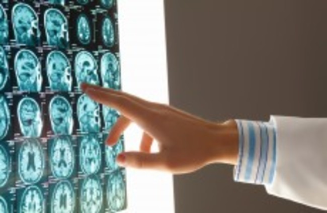
A NEW BRAIN scan technique may be able to tell whether a patient in a vegetative state has the potential to recover consciousness.

An article published in The Lancet journal outlines how traditional bedside examinations of patients in different states of unconsciousness lead to misdiagnosis in 40 per cent of cases.

The examinations are used to determine whether patients are in a minimally conscious state, in which there is some evidence of awareness and response to stimuli, or are in a vegetative state also known as unresponsive wakefulness syndrome.

Patients with the latter syndrome have a much lower chance of recovery and the new imagery known positron emission tomography (PET) as could help doctors decide on treatment.

“Our findings suggest that PET imaging can reveal cognitive processes that aren’t visible through traditional bedside tests, and could substantially complement standard behavioural assessments to identify unresponsive or “vegetative” patients who have the potential for long-term recovery”, says study leader Professor Steven Laureys from the University of Liége in Belgium.

The study used the new imagery technology during the course of mental imagery tasks that were being undertaken by the 126 patients who took part.

Jamie Sleigh of the University of Auckland explains how ineffective the current methods of examination can be in some cases:

In patients with substantial cerebral oedema [swelling of the brain], prediction of outcome on the basis of standard clinical examination and structural brain imaging is probably little better than flipping a coin.

One of the drawbacks of the new imagery however is that it may be difficult to roll out in hospitals on a wider basis. The study was carried out in a specialist unit that focuses on the neuroimaging of consciousness disorders and therefore had a level of expertise and equipment that would not be present in most clinics.

Read: Apps from Irish start-up to provide home therapy for stroke sufferers >

Read: New research shows brain tumours can be killed with anti-nausea drug >

Your Voice
Readers Comments
3
    Submit a report
    Please help us understand how this comment violates our community guidelines.
    Thank you for the feedback
    Your feedback has been sent to our team for review.