Sign in. It’s quick, free and it’s up to you.
An account is an optional way to support the work we do. Find out more.
Sign in. It’s quick, free and it’s up to you.
An account is an optional way to support the work we do. Find out more.
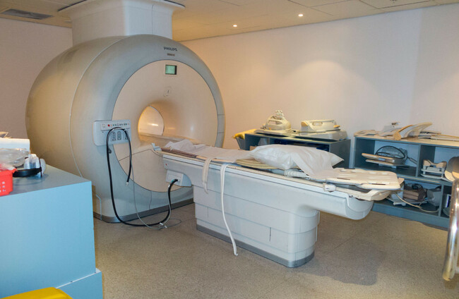

IN MY DAY job, I see the impact of back pain on patients every day. I work as a Clinical Specialist Physiotherapist at the Mater Hospital as part of a novel integrated care pathway for managing low back pain across the hospital and its community partners in Dublin city.
Many of the patients I meet have already had an MRI scan or some other form of imaging done. They often arrive with the results of their scan, expecting that it reveals the cause of their pain. The expectation that if my back hurts, there must be something structurally wrong that can be seen on a scan has become commonplace.
We live in a fast-paced, instant-gratification culture, and when pain or injury finds its way into our lives, having a scan can seem like the most reliable and necessary way to establish a diagnosis and get the symptoms “fixed”.
Yet when I assess these people clinically, the picture is often much more complicated and nuanced. Most people with back pain do not have a single structural abnormality that neatly explains their symptoms, and many of the changes seen on spinal MRI scans are also common findings in people who have no pain and can be linked to the ageing process.
This raises an important question about the culture of imaging in healthcare today: have we become too focused on finding something on a scan, rather than listening and understanding the person sitting in front of us and the importance of a comprehensive clinical physical examination?
If you develop low back pain, one of the first things you may think is I will go to my GP because ‘I need an MRI’. It is an understandable reaction. Back pain can be worrying and when severe, very debilitating. It can stop you doing the things you normally do and make daily activities such as sitting, driving, putting your shoes and socks on difficult.
However, bouts of back pain are perfectly normal. Most episodes of lower back pain are not caused by a serious underlying condition but occur as a result of a particular activity or repeated movement and will improve with time.
Yet, for some reason, people seem to interpret low back pain very differently from pain elsewhere in the body. Often people with knee pain may hobble around and think, ‘I’ve probably done something to it. It will settle.’ But when people develop back pain there can be an immediate sense that something much more serious is going on.

Perhaps back pain is associated with the spine column and spinal cord, and there is a perception that this is more serious than pain elsewhere in the body. Back pain, like any other musculoskeletal pain, is a normal part of life. It can flare up, it can be painful and limiting, and then it can settle. Evidence shows that most people with low back pain experience substantial improvement within the first six weeks, with the greatest improvements occurring early on.
Because technology can now produce extraordinarily detailed images of the inside of the body, it is understandable that when we have pain, we want to see what is going on. An MRI also called a Magnetic Resonance Image can feel like the most reliable way of finding an answer. An MRI is exceptionally good at showing anatomical structures. The challenge is understanding which of those findings, if any, are actually relevant and contributing to the person’s pain.
To explore this further, I spoke with consultant spine surgeon Mr Marcus Timlin, Clinical Advisor for the National Integrated Low Back Pain Pathway. According to Mr Timlin, the more important question is not simply whether an MRI can be done but whether it is required, and if so, what are we expecting it to tell us?
For many people presenting with lower back pain, an immediate scan is not necessary. Mr Timlin explains that there are certain features that patients present with that would make him more concerned about what is causing these symptoms including severe, unremitting pain that keeps someone awake at night, a history of fever, shivering or sweats, a history of cancer, or pain that continues rather than settling. On the other hand, someone who has developed back pain over the last few weeks, particularly pain that is related to movement or activity, may not need an MRI at all.
Evidence-based clinical guidelines for the management of low back pain recommend relative rest, i.e. avoiding activities that aggravate your symptoms, exercise modification, physiotherapy and anti-inflammatories as required These episodes will often improve quickly over a period of 2 – 4 weeks.
This can be difficult for people to hear. When something hurts, we want to know what we have done, and more importantly, what we need to do to make it better. We have become accustomed to getting answers quickly and accepting injury and the need to modify our lives in the short term can be hard.
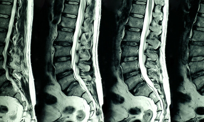
Mr Timlin explained how often he sees changes such as disc bulges and degenerative changes on MRI scans in people who have little or no back pain. He explained that changes in the spine become increasingly common as we get older, even in people who are not experiencing pain. This is important because it means that finding something on an MRI does not necessarily mean that we have found the cause of the pain.
Research supports this, showing that changes such as disc degeneration and disc bulging become increasingly common with age in people who have no symptoms. Disc degeneration, for example, was seen in 37% of people aged 20 and 96% of people aged 80, while disc bulges ranged from 30% at age 20 to 84% at age 80. These figures are striking because they show us that the spine naturally changes as we get older. Not every change is a disease, and not every abnormality seen on an MRI is the source of pain.
I echo these thoughts from my own clinical experience. Pain is much more complex than what we see on a scan. I often explain to patients that their MRI is a picture of their spine at one moment in time. It does not show how they feel, how stressed they are, how fit or unfit they are, how strong or stiff they are, or how they move. All of these factors can influence a person’s experience of pain and their recovery.
The good news is that these are things we can change. Through exercise, physiotherapy and guided support, we can help people build strength, improve their movement and confidence, and gradually return to the activities that matter to them. When we focus less on trying to find the perfect explanation of pain on a scan and more on the things we can actually change, people can begin to recover and get back to living the life they want.
So when do we know we actually need a scan? This is where the clinical assessment becomes so important. Mr Timlin explained the importance of the patient’s story. When did the pain start, where it travels, what makes it worse and what relieves it. This information is combined with a detailed clinical examination of the nerves. The decision of whether an MRI scan is needed is then considered in the context of all available information. If an MRI shows a disc bulge, the important question is whether that finding fits the symptoms and examination. As Mr Timlin explains, “This allows us to ascertain what is relevant and causing symptoms.”

Mr Timlin went on to discuss MRI reports and how when reading a report, “you might imagine there are a lot of bad/serious things going on or the ‘spine is wearing out or nerves badly compressed’.” Therefore an MRI cannot be interpreted in isolation. The findings need to be considered alongside a person’s symptoms and clinical examination to determine what is actually relevant.
Sometimes the words used in an MRI report can be more frightening than reassuring. “Degenerative changes”, “disc protrusion”, “nerve compression”, “wear and tear”. For a patient, these phrases can sound like a diagnosis of a damaged spine. More often than not, I see people who have had a scan related to their back pain, however, they have never sat with a clinician to explain the findings and review the images of their scan in detail. They usually receive a phone call from their GP to go through the report and are left with more questions than answers.
The problem then, is not on the technology but on the reliance of it as the first point of contact for a patient presenting with low back pain. This can often give what is seen on a scan more meaning than it deserves. Mr Timlin points out that “pain is personal” and everyone senses pain differently and that how we think and feel influences pain levels. He also notes that pain may come from muscles, ligaments, tendons, joints which can cause back strain but are not necessarily obvious on an MRI. That does not make the pain imaginary. Pain is real, even when there is no single abnormality on a scan that neatly explains it.
This is where early access to seeing a physiotherapist can make a significant difference in helping people, firstly to understand their pain, and then to support them in managing their symptoms through gentle exercises, stretches and activity modification. This is important because in this time of advancing technology and AI within healthcare, it would be easy to confuse finding something on a scan with finding the actual cause of a person’s pain.
There is also the question of what happens after someone leaves a consultation be that with the GP or another healthcare professional with an MRI report in their hands. If they have been told they have degeneration or a disc bulge, they may begin to think differently about their back. They may become worried about bending, stop exercising or become afraid that normal movement is damaging their spine. The finding on the scan can become the explanation for everything. Physiotherapists working with people with low back pain often want patients to push, pull, lift and carry. We want them to bend their back and exercise. To focus on strength and mobility and often do more than there are currently doing. This is often the opposite of what people with back pain do or are told to do.

This is not an argument for stopping radiological investigations, it is one for the appropriate use of technology within this patient group. There are circumstances where it is essential and can provide extremely important information. The question is whether the findings are clinically relevant and whether the scan will actually change their management plan. Perhaps the most useful question for patients to ask their relevant healthcare professional is therefore not “Can I have an MRI?” but “Would an MRI change what we do next?”
If the answer is yes, imaging may be an important part of the next step and onward referral. If the answer is no, there may be little benefit in finding abnormalities that are likely to be incidental. Perhaps the simplest message is also the most important. Not everyone with back pain needs an MRI.
And when someone does have an MRI, not everything that appears on the scan is necessarily the cause of their pain. The scan needs to be interpreted alongside the history and examination. As Mr Timlin puts it, the findings “must be relevant to their symptoms.”
Stephen O’Rourke is a Chartered Clinical Specialist Musculoskeletal Physiotherapist at the Mater University Hospital, Dublin, with a specialist focus on spinal care and low back pain. He is also a guest lecturer at the Royal College of Surgeons in Ireland and is a health contributor to TheJournal.ie.
To embed this post, copy the code below on your site
















have your say